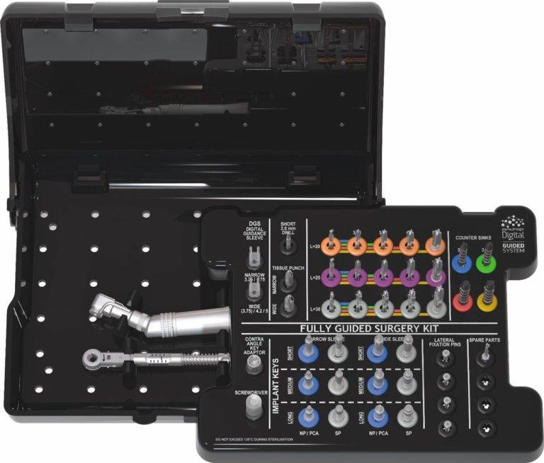
WHAT IS REQUIRED FOR DUAL SCAN PROTOCOL?
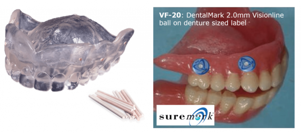
A full denture /radiographic guide which is radiolucent & has 6 -8 radiopaque markers (e.g. Suremark or Gutta Percha)
WHAT IS REQUIRED FOR DUAL SCAN PROTOCOL?
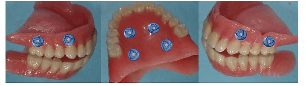
- Place 4 markers in the palate area
- Place 4 markers on the buccal side between the necks of the denture teeth & the edge of the flange
WHAT IS REQUIRED FOR DUAL SCAN PROTOCOL?
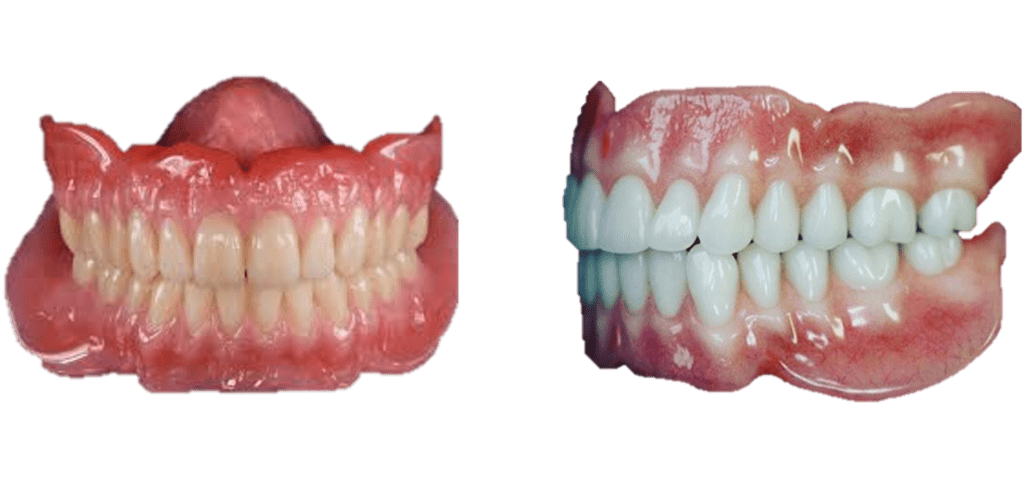
- The tooth set-up should be ideal and represent the final desired result.
- The correct vertical dimension.
- In the case of an upper denture it must have full palatal coverage.
WHAT IS REQUIRED FOR DUAL SCAN PROTOCOL?
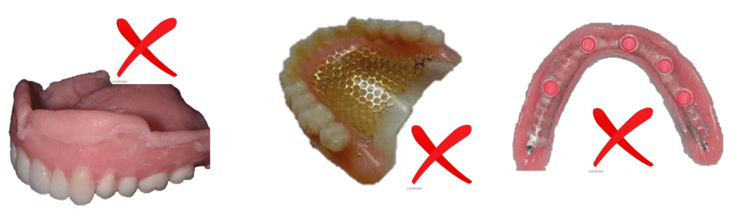
The full denture /radiographic guide must be stable & close fitting to the soft tissue & have no metal components:
e.g. wire mesh/plate, metallic crowns, wire clasps
It is recommended that the denture has a hard reline.
WHAT IS REQUIRED FOR DUAL SCAN PROTOCOL?
- The geometry of radiographic guide is transferred to the Surgical Template
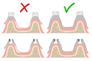
- It is essential that the radiographic guide/denture is strong, rigid, reaches the full depth of the sulcus and is stable on the mucosa
COLLECTION OF DIGITAL INFORMATION FOR FULLY EDENTULOUS CASES
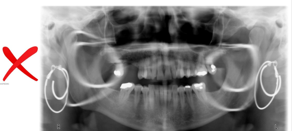
- Before the CBCT is taken the patient should remove all jewelry & removable appliances with metal components
- Please note : when the patient has fixed metal prosthetic components in his/her mouth, this will affect the quality of the CT & may cause unwanted scatter.
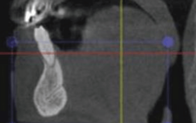
CBCT DUAL SCAN PROTOCOL FOR FULLY EDENTULOUS
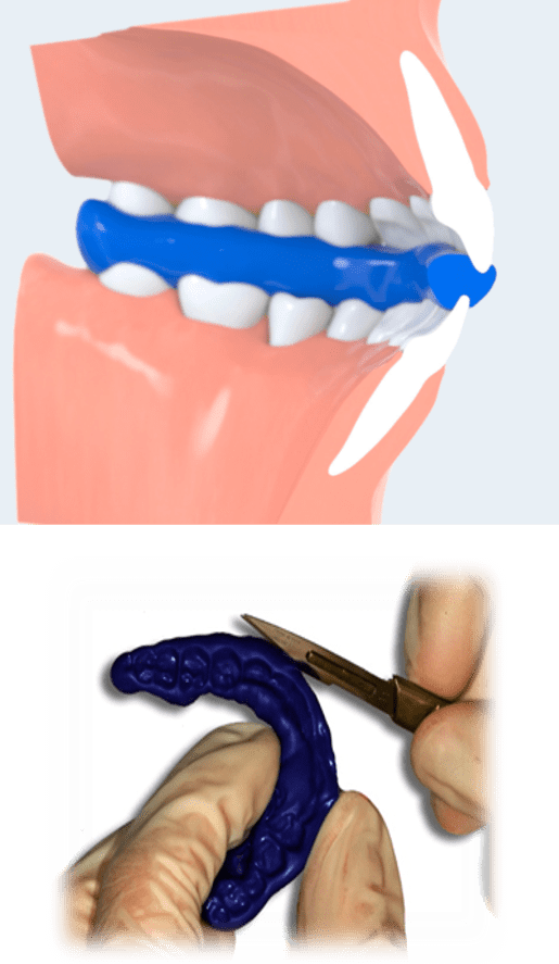
- It is recommended to prepare a bite index to stabilize & separate the jaws when scanning the patient only!
- Apply radiolucent material such as “aquasil” on to the occlusal surface. The patient should not close fully maintaining a small gap of 2-3mm. Trim away excess material.
CBCT DUAL SCAN PROTOCOL FOR FULLY EDENTULOUS
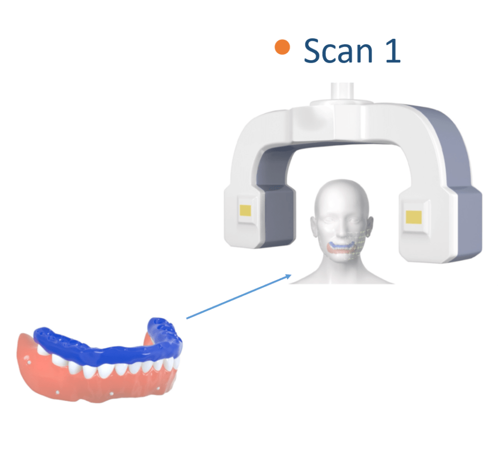
- Scan the patient wearing the radiolucent denture or scan prosthesis with 8 scan markers
- To stabilize the denture it is recommended to use a radiolucent bite index
CBCT DUAL SCAN PROTOCOL FOR FULLY EDENTULOUS
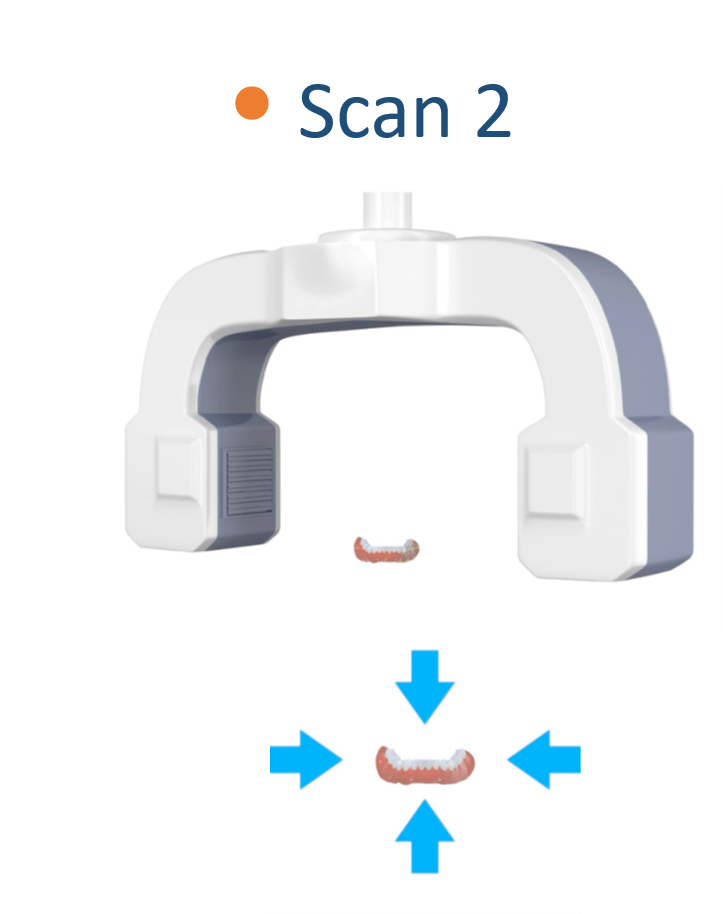
- Scan the prosthesis alone in the same orientation as taken in scan 1. Ensure to remove the bite index!
- The material used to support the prosthesis must be more radiolucent than the prosthesis itself.
e.g. polyurethane & polyurethane foam materials
CBCT DUAL SCAN PROTOCOL FOR FULLY EDENTULOUS
Export the raw dicom axial files only
Prepare two separate folders
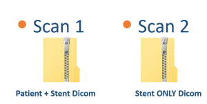
- Patient’s name must be present in the Dicom files
- Variable slice thickness is not allowed
- Gantry tilt 0ᵒ
- Only axial images required
- The scans must be saved as “Dicom” files
- The files should be “zipped” before upload

Mr. Philip Segal, MDT
PALTOP CAD/CAM MANAGER