COLLECTION OF DIGITAL INFORMATION FOR PARTIALLY EDENTULOUS CASES
- Before the CBCT is taken patient should remove all jewelry & removable appliances with metal components
- Please note : when the patient has fixed metal prosthetic compoents in his/her mouth, this will affect the quality of the CT & may cause unwanted scatter.
- Temporary acrylic bridges should be without metal strengtheners
MAXILLA:
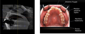
- The scan must include ALL the teeth, the entire hard palate and the tuberosity’s.
- In addition it should include some slices of the lower jaw for reference.
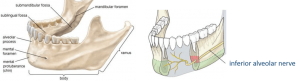
MANDIBLE:
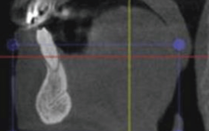
- The scan must include ALL the teeth, the ramus & the inferior alveolar nerve
- In addition it should include some slices of the upper jaw for reference.
- The digital scans should include both maxilla and mandible jaws together with the correct occlusion.
- IMPORTANT: the accuracy of the surgical guide is dependent on the digital models
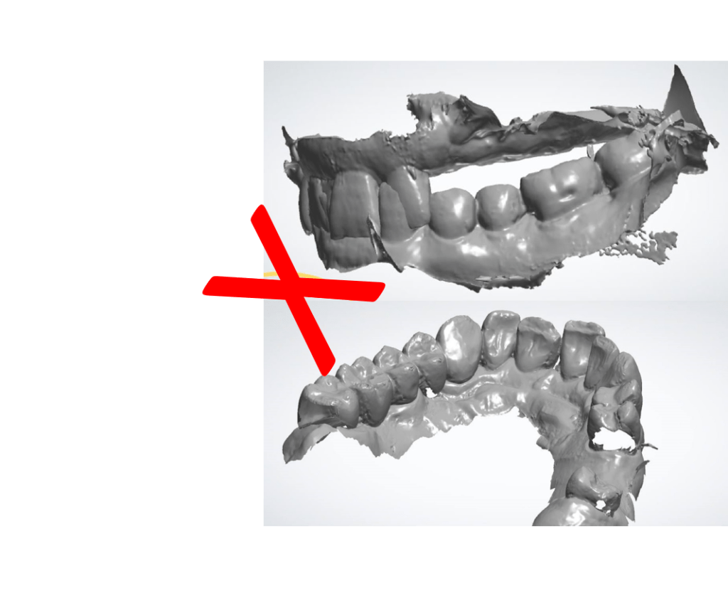
- Both the Dicom files and the Intra Oral Scans or Plaster model scans must match each other.
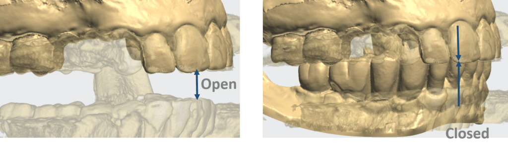
- The CT scan should be taken in open occlusion:
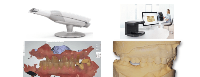
- Clinical images offer valuable information
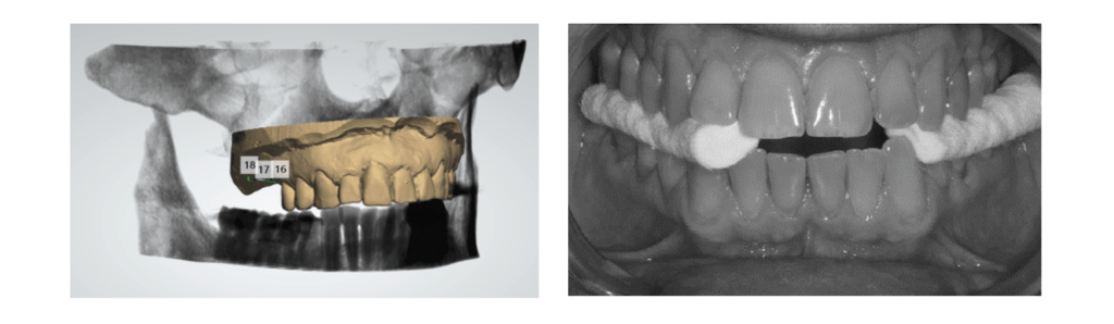
- The CT scan should be taken in open occlusion:
- Intra Oral Scan or plaster model scan in closed occlusion:
CBCT DUAL SCAN PROTOCOL FOR PARTIALLY EDENTULOUS
Export the raw dicom axial files only
- Patient’s name must be present in the Dicom files
- Variable slice thickness is not allowed
- Gantry tilt 0ᵒ
- Only axial images required
- The scans must be saved as “Dicom” files
- The files should be “zipped” before upload
Philip Segal graduated from the Manchester Dental Hospital UK gaining a diploma with distinction in Dental technology. From 1980 to 1999 he was a partner in a leading dental laboratory in Tel Aviv specializing in the field of cad/cam & aesthetic dentistry. From 1997 he served as a consultant for NobelBiocare -Israel, offering training & education to clinicians and dental technicians on Procera cad/cam technologies and All-on-4 technique. Since 2005 he has dedicated his time exclusively to the field of computer-guided surgery functioning as a mentor and instructor for NobelGuide, SIMPLANT & 3Shape Implant Studio softwares. He has played a leading role in offering planning services and 3d treatment proposals with the aid of computer-guided software and has given many lectures and courses both nationally & internationally, illustrating the use of 3D diagnostics, treatment planning & CAD/CAM technologies.
Mr. Philip Segal, MDT
PALTOP CAD/CAM MANAGER